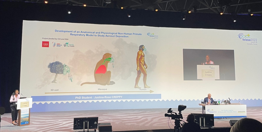

Justina Creppy was a laureate of the first edition of the international competition "My 3Rs in 180 seconds", which took place in June at the FELASA congress in Marseille. The prize, sponsored by Sanofi, was presented by the Director of the GIS FC3R, Athanassia Sotiropoulos.
A 3D model of the airway
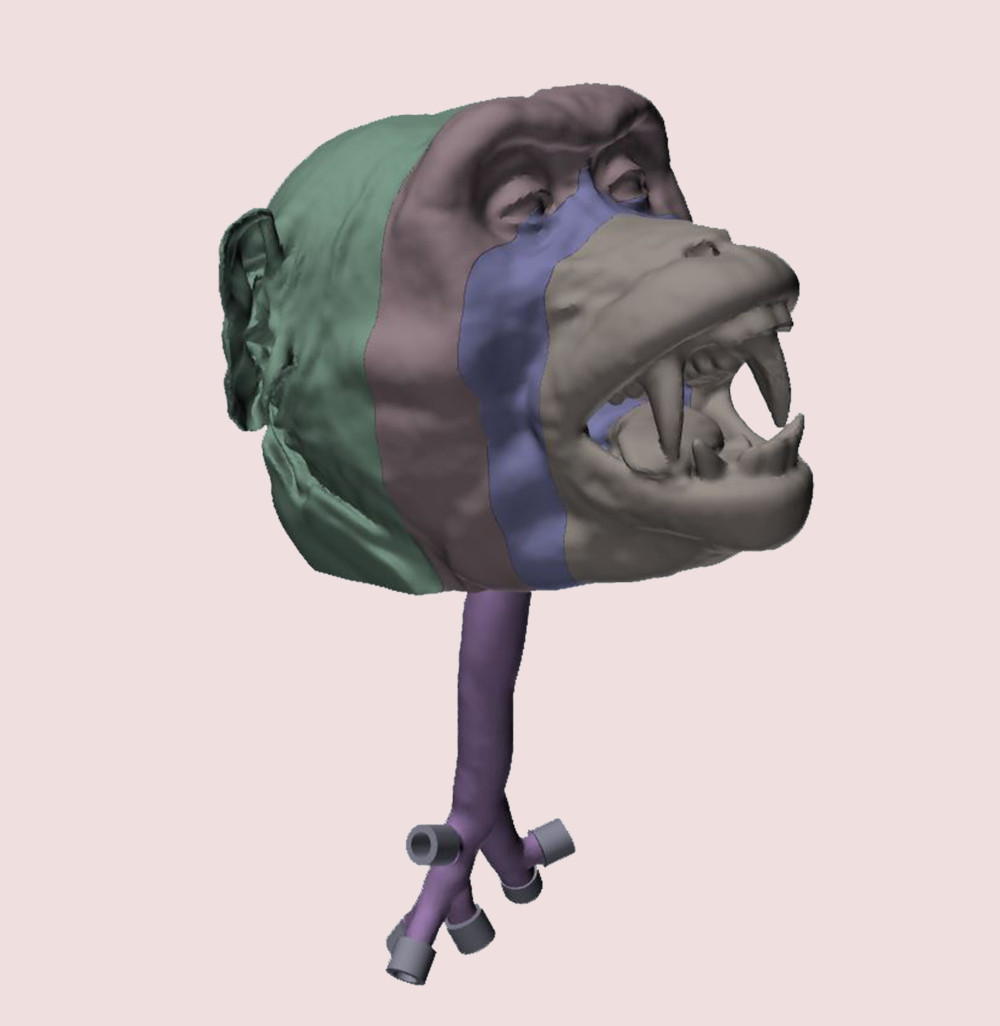
Justina Creppy is carrying out her thesis under the joint supervision of the CEA/IDMIT (under the direction of Dr. F. Ducancel), and the CEPR of the Faculty of Medicine of Tours (with Dr. L. Vecellio). Her project, funded by the AID (Agence Innovation Défense), consists in studying respiratory pathologies - and more particularly aerosol dispersion - through the development of an anatomical and physiological respiratory model of a non-human primate. She performed CT imaging of her model, a macaque Macacca fascicularis. A model was 3D-printed based on this imaging, in a waterproof, non-porous, transparent and washable material. For convenience, the model was not printed in one block, but detailed in 4 different modules (modeled below in gray, blue, purple and green) that fit together.

A realistic model to Refine
The model developed by Justina's team presents major improvements compared to already existing models (Kesavan et al., 2020), which facilitate its use while considerably widening the field of possible applications. In the quest for refinement, a research effort was thus focused on the realism of the external part of the model : the head of the monkey. This part is indeed important for the placement of the nebulization mask. The different adjustments can be optimized thanks to the plastic model, and thus save precious time during the experiments with the animal, while ensuring the most adapted conditions to its physionomy. This also allows the nebulizer users to be trained in all the technical gestures (mask placement, machine connection...) without unnecessary stress on the animal.
The addition of the lower airway to Replace partially
But the biggest innovation of this project is the addition of the lower airways. Justina and her team scanned and then 3D printed the trachea down to the third bronchial divisions of the simian model. The size of the animal, a male macaque weighing nearly 6 kg, made it possible to access these fine structures, which could be connected to a pump to mimic the animal's respiratory movements. With this anatomical and physiological model, it is possible to study the behavior of aerosolized particles in the model, the locations and the pattern in which they are deposited in the upper and lower airways.

Prospects in research and human medicine
Chemical, bacterial and viral aerosols can thus be traced by fluorescence or radioactivity monitoring (Leclerc et al., 2014), which presents interesting therapeutic perspectives for many respiratory infections. In particular, this model is used in Covid-19 research : it improves our understanding of the behavior of the virus after inhalation, allows to identify its anatomical targets and thus to predict which aerosols or nebulization methods could be effective in therapy. The 3D model also allows the optimization of aerosol delivery systems (masks, nasal cannulas, etc.) without the use of live animals (Williams and Suman 2022). In comparative study with animals, the 3D printed model showed good predictivity of the total amount of aerosol deposited within the upper airways (Le Guellec et al., 2021)(Kelly et al., 2005), but an overestimation of the deposition in the lower airways, due to the absence of the rest of the lung.
While this is a good model, respectful of the 3Rs, which is proving its worth in the research fields concerned, it does not yet dispense from complementary experiments on animal models.
